Introduction: Quantitative EEG in ASD as a Window into Neurodevelopment
Quantitative EEG in ASD (Autism Spectrum Disorder) provides a unique lens to observe and understand the neurodevelopmental deviations that characterize this complex condition. Brain development in children is a dynamic and intricate process, marked by changes in synaptic density, neural connectivity, and electrical activity. Electroencephalography (EEG), and specifically quantitative EEG (qEEG), offers a window into this unfolding neural landscape by measuring and analyzing brainwave patterns across development. In children with Autism Spectrum Disorder (ASD), these electrical signatures often diverge from typical developmental patterns, providing critical insights into the underlying neurobiology of the disorder. This article explores the scientific understanding of EEG features in ASD, the developmental trajectory of brainwaves, their clinical relevance, and implications for therapeutic intervention.
Why qEEG? Understanding the Value of Quantitative EEG
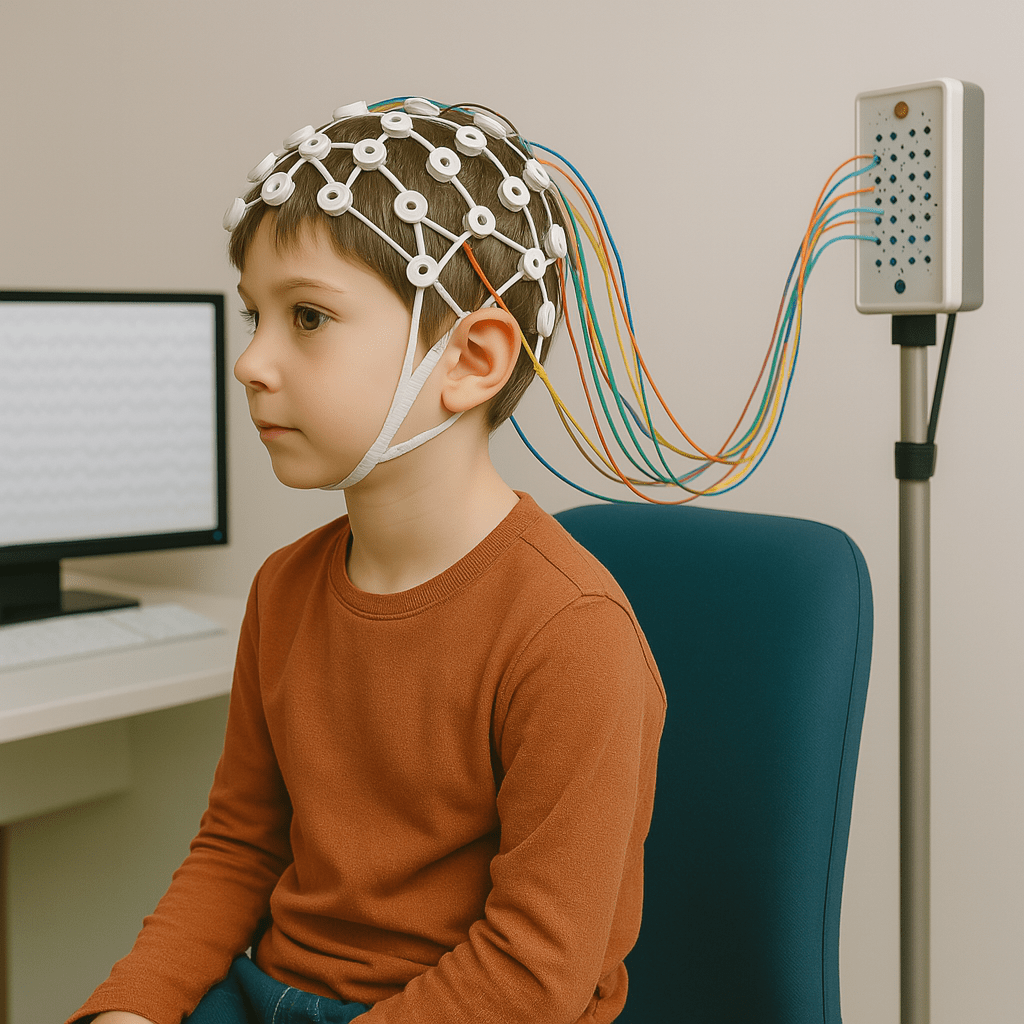
To understand the neurodevelopmental underpinnings of autism, clinicians and researchers increasingly turn to quantitative EEG (qEEG) rather than conventional EEG. While traditional EEG is designed to detect overt abnormalities like seizures or epileptiform discharges, qEEG allows for a comprehensive, statistical evaluation of brainwave dynamics—analyzing power, coherence, phase relationships, and asymmetry across multiple cortical areas.
This quantitative approach is particularly relevant in ASD, where standard imaging often fails to capture functional brain differences. Quantitative EEG in ASD helps identify subtle disruptions in neural communication and oscillatory regulation that contribute to sensory dysregulation, attention deficits, and social impairment. It enables individualized comparison to large normative databases, which helps pinpoint deviations specific to the ASD neurotype.
qEEG provides:
- Detailed frequency band power distribution (e.g., elevated delta, reduced alpha)
- Inter-regional coherence metrics, revealing network synchrony or disconnection
- Hemispheric asymmetry patterns, associated with language and emotional regulation issues
In ASD, these metrics often highlight persistent slow-wave activity, reduced frontal alpha power, and long-range hypoconnectivity. These features make EEG in ASD an invaluable tool—not just for diagnosis, but for monitoring neurodevelopment and guiding targeted interventions like MeRT and neurofeedback.
Normal Brainwave Development: Characteristic of Quantitative EEG in Typical Children

In typically developing children, the brainwave patterns evolve systematically with age, following a trajectory of increasing neural efficiency. The early years are characterized by the dominance of slower oscillations (delta and theta), which are indicative of immature cortical circuits. As the child matures, there’s a notable shift toward faster frequencies (alpha and beta), reflecting improved cortical organization, synaptic pruning, and functional specialization.
This transformation is especially prominent in the context of cortical development. As the cerebral cortex undergoes structural and functional maturation, there is a reduction in slow-wave activity and an increase in faster waveforms, particularly in the alpha and beta bands. These changes are closely tied to enhanced sensory processing, attentional regulation, and executive function. Increased utilization of faster frequencies, especially alpha activity during wakeful rest, is viewed as a marker of efficient top-down modulation in typically developing children.

- Delta (0.5–4 Hz): Prominent in infancy; associated with deep sleep and early neural plasticity.
- Theta (4–8 Hz): Peaks during preschool years; linked to language acquisition and emotional regulation.
- Alpha (8–12 Hz): Emerges during early childhood; supports attentional control and sensory integration.
- Beta (12–25 Hz): Increases into adolescence; correlates with executive functioning and problem-solving.
The transition from slow to fast wave dominance is not merely maturational but reflects ongoing synaptic pruning, myelination, and cortical specialization.
Neurodevelopmental Divergence and Scientific Evidence: Quantitative EEG in ASD Abnormalities
The developmental trajectory of children with Autism Spectrum Disorder (ASD) diverges significantly from typical patterns, particularly in the realm of brainwave activity as captured by quantitative EEG (qEEG). EEG in ASD has uncovered a range of consistent deviations from normative brain maturation, and these anomalies are increasingly supported by a growing body of scientific literature.
One of the most commonly reported abnormalities in ASD is the elevation of delta (0.5–4 Hz) and theta (4–8 Hz) power. These slow-wave frequencies are typically dominant during early infancy, when the cerebral cortex is still immature. In neurotypical children, these slow waves gradually diminish with age as the brain undergoes synaptic pruning and increased myelination. However, in children with ASD, these slow rhythms often persist beyond expected developmental windows, suggesting delayed cortical maturation and impaired thalamocortical regulation.
Another prominent finding in EEG in ASD research is reduced alpha power (8–12 Hz) in global region of the brain. Alpha waves are strongly associated with resting-state modulation, sensory filtering, and attentional control. In children with ASD, diminished alpha power may point to deficits in sensory gating and cognitive integration. Furthermore, alpha desynchronization, which typically occurs during cognitive engagement in neurotypical individuals, appears blunted in ASD, reflecting challenges in task-related neural flexibility.
Frontal and temporal asymmetries are also observed in many studies of EEG in ASD. These asymmetries suggest disrupted hemispheric specialization—particularly affecting regions responsible for language comprehension, social reasoning, and emotional processing. For instance, a dominance of slow-wave activity in the left temporal region has been linked with expressive language delays and phonological processing deficits in ASD populations.
Connectivity metrics derived from qEEG, such as coherence and phase-locking values, offer further insight into network-level dysfunction in ASD. Decreased coherence between frontal and posterior regions is frequently reported. This long-range underconnectivity undermines the integration of top-down and bottom-up information streams, leading to difficulties in planning, executive function, and social cognition.
These patterns are not merely theoretical—they are backed by robust scientific evidence:
- Coben et al. (2008) identified elevated delta and theta power alongside markedly reduced inter-hemispheric coherence in children with ASD. The study found these abnormalities to be most pronounced in frontal and central regions, suggesting impaired communication between brain hemispheres and delayed cortical maturation. These findings provide strong electrophysiological evidence of functional disconnection, particularly affecting executive function and integrative processing networks. Their data further emphasized that such coherence deficits are not randomly distributed but are organized patterns consistent with ASD symptomatology.
- Wang et al. (2013) showed a characteristic ‘U-shaped’ EEG power profile in ASD, marked by elevated delta/theta power, reduced alpha activity, and increased beta/gamma power. This pattern was most pronounced in the frontal and parietal regions, suggesting a disruption in both low- and high-frequency regulation. They also noted hemispheric asymmetry, particularly greater slow-wave power in the left hemisphere, and significantly decreased coherence between frontal and posterior brain regions. These abnormalities were closely associated with impairments in communication and cognitive integration.
- Murias et al. (2007) observed EEG in ASD individuals showing increased local coherence in the delta and theta bands—particularly in the left frontal–temporal regions—alongside reduced global coherence in the low-alpha band. This specific EEG in ASD profile supports the hypothesis of dual network disruption: excessive local connectivity and insufficient long-range integration. These patterns reflect inefficiencies in information coordination and are especially evident during tasks that demand social attention and executive functioning.
- Mathewson et al. (2012) examined EEG in ASD adults and found significantly reduced alpha suppression during eyes-open conditions, along with lower alpha power and decreased posterior coherence. These EEG in ASD abnormalities were associated with heightened perceptual detail focus and reduced neural synchrony, supporting theories of automatic detail processing and impaired integrative attention in autism.
Recent advancements also point to the predictive and diagnostic potential of EEG in ASD. Machine learning algorithms trained on qEEG data have achieved high sensitivity and specificity in distinguishing children with ASD from neurotypical peers. These tools could one day serve as adjuncts to behavioral assessments, especially for younger children or those with limited verbal communication.
Interestingly, EEG abnormalities can often be detected early in life, sometimes even before core behavioral symptoms of ASD fully manifest. This raises the possibility of using qEEG not only as a diagnostic adjunct but as a tool for early identification and preemptive intervention. Studies have shown that earlier therapeutic engagement leads to better long-term outcomes, especially when interventions are tailored to an individual’s neurophysiological profile.
Cross-cultural studies from Asia, Europe, and North America reinforce the universality of these EEG in ASD findings. Despite differences in language, culture, and healthcare infrastructure, consistent patterns of elevated slow-wave activity, reduced alpha/beta power, and network underconnectivity have been observed. This suggests that the qEEG abnormalities in ASD reflect core neurodevelopmental mechanisms rather than environmental variance.
The integration of EEG in ASD research continues to yield critical insights into the biological basis of autism. The converging evidence from neurophysiological data, behavioral correlations, and computational modeling indicates that qEEG is not only a valuable research tool but a promising clinical adjunct. As our understanding deepens, EEG-based markers may help usher in a new era of precision medicine in autism care, guiding early diagnosis, individualized therapy, and continuous monitoring of neural progress.
Clinical Implications of EEG in ASD
The atypical brainwave signatures observed through EEG in ASD are not merely academic—they provide actionable insights with direct relevance to clinical decision-making and patient care. Quantitative EEG (qEEG) enables clinicians to translate subtle deviations in brainwave patterns into targeted intervention strategies tailored to an individual child’s neurological profile.
For instance:
- Elevated Delta/Theta Activity is commonly associated with delayed speech development, attentional dysregulation, and difficulties with emotion regulation. Children with prominent slow-wave dominance may appear lethargic, distractible, or overwhelmed in multi-sensory environments.
- Reduced Alpha Power, particularly in posterior and frontal areas, correlates with challenges in sensory integration, resting-state modulation, and social disengagement. This EEG in ASD profile often aligns with behaviors such as withdrawal, hyperresponsivity to sensory stimuli, and difficulty with transitions.
- Frontal Asymmetry, especially when left-dominant delta or theta power is present, has been linked to expressive language delays and behavioral rigidity. Understanding hemispheric differences helps clinicians identify imbalances in emotional and linguistic processing.
- Network Hypoconnectivity, reflected as decreased coherence across frontal-posterior or interhemispheric regions, indicates impairments in top-down regulation. This may manifest as poor problem-solving, weak impulse control, and limited theory of mind abilities.
The value of EEG in ASD also extends to treatment monitoring. Repeating qEEG after a cycle of therapy—whether behavioral, neurofeedback, or pharmacological—can reveal shifts in coherence or frequency distribution that precede or parallel behavioral change. For example, a rise in frontal alpha coherence may signal improving executive function even before the child demonstrates it overtly in everyday settings.
Clinicians are increasingly recognizing that observable behavior may only be the surface expression of deeper network dysfunction. qEEG offers a “functional biomarker” to illuminate the underlying brain mechanisms, helping bridge the gap between symptom and system. This is especially useful in cases where standardized behavioral assessments fall short—such as in minimally verbal children or those with comorbid conditions.
In the future, EEG in ASD may also play a role in treatment stratification, helping determine which interventions are likely to be most effective for a particular neurophysiological subtype. As machine learning and large normative datasets evolve, the potential to classify EEG in ASD into clinically meaningful endophenotypes could revolutionize individualized care.
Ultimately, integrating qEEG into clinical workflows offers a powerful opportunity to not just observe, but to act—enabling clinicians to deliver brain-based, personalized care to children with ASD.
Therapeutic Interventions Informed by Quantitative EEG in ASD
Emerging neurotherapies informed by EEG in ASD aim to directly address the atypical neural oscillations and connectivity patterns that underlie behavioral symptoms. These interventions represent a paradigm shift in autism treatment—from broad-spectrum behavioral approaches to targeted neuromodulatory techniques grounded in brain physiology.
- Neurofeedback (NFB): Neurofeedback therapy involves using real-time EEG to help individuals with ASD learn to self-regulate specific brainwave patterns. For example, children with excessive theta activity and deficient beta power may be trained to reduce theta while enhancing beta, thereby improving sustained attention, working memory, and impulse control. Multiple studies have demonstrated that EEG in ASD-guided neurofeedback can reduce hyperactivity and improve social behavior, particularly when training protocols are individualized based on qEEG assessments.
- MeRT (Magnetic e-Resonance Therapy, EEG guided rTMS): MeRT represents an advanced and individualized form of EEG-guided brain stimulation. It begins with a detailed qEEG analysis to identify each child’s unique neurophysiological markers—such as dominant frequency imbalances, asymmetrical connectivity, or delayed alpha rhythm maturation. Using this information, clinicians develop a personalized magnetic stimulation protocol targeting areas of underconnectivity or desynchronization. Unlike conventional TMS, MeRT leverages EEG in ASD to time and localize stimulation with precision, promoting realignment of neural circuits. Clinical evidence shows that MeRT can lead to improvements in multiple functional domains, including receptive and expressive language, social reciprocity, eye contact, emotional regulation, and sleep quality. It is particularly promising for children with ASD who exhibit frontal alpha deficiency, poor interhemispheric coherence, or prolonged theta dominance. By supporting neuroplastic reorganization in these dysfunctional networks, MeRT facilitates the brain’s return to a more optimal state of synchronization and integration. As a noninvasive, qEEG-informed intervention, MeRT offers a compelling model for personalized, circuit-based therapy in ASD.
- At BraintreatmentCenter Seoul, we have applied MeRT extensively in children with neurodevelopmental conditions, particularly Autism Spectrum Disorder. Based on our clinical experience, MeRT not only induces short-term changes in brain function but also promotes sustained improvements across critical developmental domains. We have observed meaningful gains in sleep quality, cognitive processing, and the regulation of sensory input. Notably, many children show improved emotional regulation, reduced sensory-seeking behaviors, and increased social interaction over the course of MeRT treatment. In several cases, caregivers have reported that these changes translate to improved quality of life at home and school. Through continuous qEEG monitoring, we tailor protocols as each child’s brain dynamics evolve, ensuring that stimulation remains precisely targeted and adaptive over time. Our observations affirm that EEG in ASD is more than a diagnostic tool—it is the foundation for transformative therapeutic planning and ongoing clinical feedback.
- Sensory Integration and Cognitive Training: Interventions targeting sensory processing or cognitive control—such as auditory integration therapy, interactive metronome training, Sensory Integration Therapy (SIT) or computerized working memory programs—can be optimized when paired with EEG in ASD data. For example, children with reduced posterior alpha power may benefit from programs that strengthen sensory gating and visual processing circuits.
- Pharmacological Personalization: While not a standalone EEG-based therapy, qEEG findings in ASD may also inform psychopharmacology. For instance, high frontal theta may suggest benefit from stimulant medications; low alpha coherence might predict poor response to SSRIs. EEG in ASD may thus guide both neuromodulation and medication decisions in an integrated care model.
EEG in ASD provides more than diagnostic insight—it enables tailored therapeutic intervention. The future of autism treatment lies in this intersection of neurotechnology, brain-based assessment, and individualized care. By mapping dysfunctional circuits and monitoring change over time, qEEG-guided therapies hold the potential to optimize outcomes and promote meaningful developmental progress in children on the autism spectrum.
Limitations and Future Directions for Quantitative EEG in ASD
While promising, qEEG in ASD still faces challenges:
- Interindividual Variability: ASD is heterogeneous; not all children exhibit identical EEG patterns.
- Artifact Sensitivity: Movement and compliance issues can limit data reliability in younger children.
- Standardization Issues: Normative databases need to be age- and region-specific to enhance diagnostic utility.
Future research should aim to integrate EEG with other modalities (fMRI, genetics), develop age-stratified norms, and refine intervention algorithms.
Conclusion: From Brainwaves to Breakthroughs
Quantitative EEG provides a powerful lens into the neurophysiological underpinnings of autism spectrum disorder. By revealing how brainwave development in ASD deviates from typical patterns, it offers both diagnostic insights and pathways for targeted intervention. As technologies advance and evidence grows, qEEG-guided therapies may well become a cornerstone in personalized care for children with ASD, transforming brain data into developmental breakthroughs.
If you have questions or would like to inquire about brain-based therapies like MeRT, feel free to contact us at: [email protected]